Endometrial Regeneration
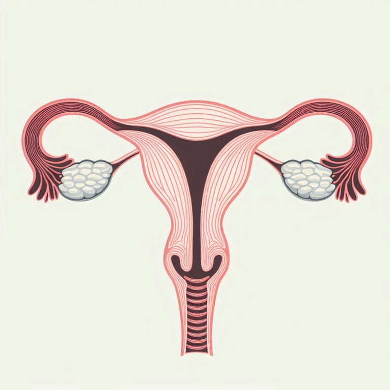
A properly functioning endometrium is a key factor for embryo implantation and pregnancy development, which we support at DeCure Medical Research.
The ability to conceive, maintain pregnancy, and give birth for a healthy 20-year-old woman is relatively low, at 20% per cycle. For a woman to become pregnant, not only gametes (egg and sperm) are needed, but also an appropriate environment in which the fertilized egg can develop. This environment is the endometrium.

The exact mechanism by which an embryo implants in the endometrium is not yet known. The most popular theory suggests that it happens through signals sent from the embryo to the endometrium and vice versa, while simultaneously suppressing the immune system. An essential element of this process is the proper reactivity of the uterine lining, which we monitor by measuring its thickness in a transvaginal ultrasound. Through the use of molecular diagnostics and the expression of 238 genes present on the endometrial surface, it has become possible to individually assess the “implantation window”.
Performing embryo transfer at the optimal moment has become the basis for increasing the effectiveness of the in vitro fertilization procedure.
Lack of endometrial growth and, consequently, lack of its reactivity results in repeated embryo implantation failures, a situation where a properly formed and healthy embryo does not implant in the uterine cavity.
Existing treatment methods include:
- hormone replacement therapy (estrogen administration)
- improving vascular flow by administering type 5 phosphodiesterase inhibitors (sildenafil) – endometrial scratching
- controlled endometrial damage to stimulate its reactivity
- surrogacy – transferring the embryo to another woman’s body (method prohibited in Poland).
The methods described above have not brought the expected therapeutic effects and are proposed with varying success as leading ways to stimulate endometrial growth. In the case of iatrogenic endometrial damage, hysteroscopy has been the standard treatment for years. Malformations within the uterus are one of the causes limiting fertility. It is estimated that they account for about 5-10% of female infertility causes. In this group, 2% to 20% of women suffer from Asherman’s syndrome or intrauterine adhesions.
MSC - Mesenchymal Stromal Cells
Mesenchymal Stem Cells
We present a breakthrough solution in tissue regeneration, based on years of research on mesenchymal stem cells (MSC). The results of our efforts have been recognized by the scientific community and published in professional literature.
Versatile Regenerative Potential
Unique Immunomodulatory Properties
Local Repair Coordination
Safe and Ethically Sourced
The term “mesenchymal stem cells” (MSC – mesenchymal stromal cells) is defined as multipotent progenitor cells with the ability to differentiate and mature into cartilage, bone, and adipose tissue cells. They serve a supportive function for other stem cells in the production of connective tissue in individual organs and form the bone marrow stroma for hematopoietic cells. They also have the ability to modulate immune system functions. According to the classification introduced by the International Society for Cell Therapy, MSCs should meet three conditions: have the ability to grow in vitro in an adherent form, possess specific expression of surface antigens such as CD13, CD44, CD90, CD73, CD105 while lacking CD14, CD11b, CD79, CD34, CD45, and HLA-DR, and be capable of differentiating into bone, cartilage, and adipose tissue.
During fetal development, MSCs are present in fetal blood (cord blood), Wharton’s jelly, perivascular area, submucosa, umbilical cord, placenta, and amniotic fluid. In contrast, clinically used MSCs taken from cord blood and Wharton’s jelly are characterized by an easily obtainable appropriate amount of material without ethical issues.
They induce an immunosuppressive effect on lymphocytes and inhibit T lymphocyte proliferation [39]. These immunological properties of these cells result in both the lack of the recipient’s immune system reaction to mesenchymal cell transplantation and the lack of allogeneic mesenchymal cells’ reaction to functioning in the recipient’s tissue system. It is believed that stem cells act as a local coordinator of tissue repair. Repair of damaged tissue occurs through the regulation of endogenous regenerative processes, not by replacing damaged tissue with de novo structures derived from mesenchymal cells. Fetal MSCs have a greater expansion potential in in vitro culture than adult mesenchymal stem cells.
Many authors believe that this is a consequence of two primary hematopoietic cell passages from the embryo to the placenta and back. The first passage, between 4 and 12 days of embryogenesis, begins through the primitive umbilical cord, and hematopoietic cells migrate from the yolk sac to the placenta. Then, during the second migration, MSCs are transferred in the opposite direction. They return from the placenta to the fetus: to the liver, and then to the bone marrow. Scientists believe that during this migration, mesenchymal cells are trapped in Wharton’s jelly, where they remain from early embryogenesis throughout the entire pregnancy until birth. WJ-MSCs can interact in the donor’s body through three independent mechanisms: differentiation into various tissue types, through an immunomodulatory effect, and through the ability to regulate external effects occurring in the MSC environment by secreting appropriate cytokines and direct contact with other cells. According to the latest scientific reports, the most important role of MSCs is considered to be local coordination of tissue repair. The result of interactions between MSCs, other cells, and tissue regeneration mediators can potentially adapt MSC signaling to the changing situation. However, it has been shown that structures created directly by descendant cells derived from MSCs are relatively rarely responsible for repairing the damaged area.
The use of MSCs in patients suffering from endometrial atrophy and Asherman’s syndrome has allowed for full restoration of the procreative function of the uterine lining. Endometrial regeneration is a permanent process, and the effectiveness of the therapy is evidenced by the qualification of patients for frozen embryo transfer obtained in the in vitro fertilization program.
The results of our efforts have been recognized by the scientific community and published in professional literature.

